Mechanical modelling of living plant cells and tissues

Mechanical forces play a key role in regulating growth, physiology and gene expression of plant cells. Plant tissues have a complex architecture, which makes it difficult to predict the internal forces acting at the cellular level. We use the latest modelling tools developed in Richard Smith lab to compute force patterns in plants and understand their biological significance.
Cellular Force Microscopy

Cellular Force Microscopy is a new micro-indentation technique dedicated to force measurements and micro-surgery in biological tissues, in particular plants. We originally developed CFM combined with light microscopy to measure the stiffness of single cells or transparent samples (Routier-Kierzkowka et al. 2012, Weber et al. 2015). We are currently working on combining CFM with confocal microscopy and 3D image analysis in MorphoGraphX.
More about Cellular Force Microscopy.
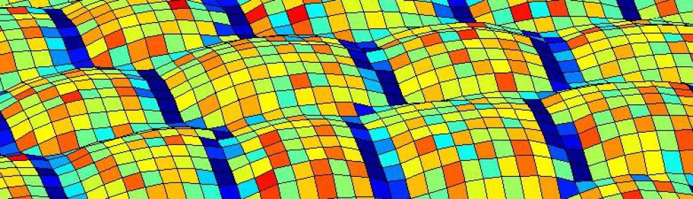
MorphoGraphX

MorphoGraphX started as a software for 3D image analysis and is now an interdisciplinary platform to quantify growth, cell shape, gene expression and mechanics.
Downloads, documentation and more about MorphoGraphX on the software website, www.morphographx.org.
Our paper about MorphoGraphX was published in eLife and awarded by the journal redaction. More about MorphoGraphX in The Node.
PAST PROJECTS:
Measuring tissue mechanics

In collaboration with the group of Angela Hay (MPI Cologne) and Alain Goriely (Oxford, UK), I measured the forces involved in one of the fastest movements occuring in plants. This project illustrates the power of a multi-disciplinary approach to explain mechanical behaviour from the cell level to the organ scale. We recently published our findings and presented them in a short video.
