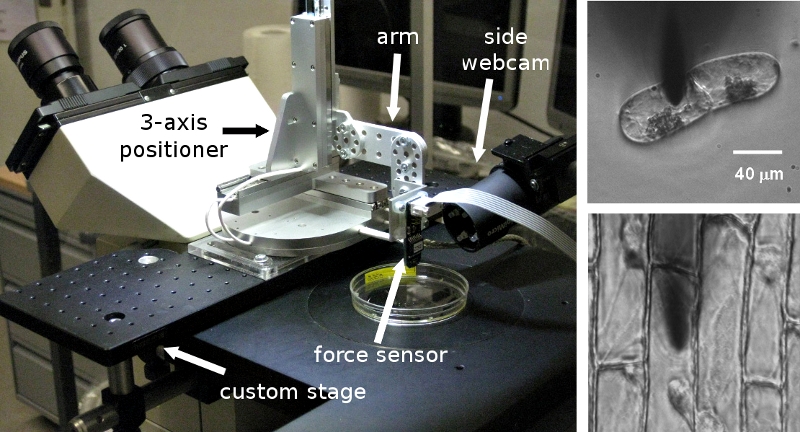
Fig2. Left: CFM on an inverted light microscope. Right: microscopy pictures showing the samples (here, BY2 cells and onion epidermis) and probe tip during the measurement.
Fig2. Left: CFM on an inverted light microscope. Right: microscopy pictures showing the samples (here, BY2 cells and onion epidermis) and probe tip during the measurement.
