
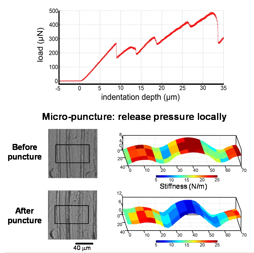
Fig4. Top: force-displacement curve obtained during cell wall puncture. Bottom: Light microcsopy images and stiffness maps of a cell before and after puncture.

Fig4. Top: force-displacement curve obtained during cell wall puncture. Bottom: Light microcsopy images and stiffness maps of a cell before and after puncture.
